
One way to understand how modern-day humans have evolved from their ancestors is to compare bone and tooth growth in people today with growth patterns
in early humans.
Scientists working in the field of human paleobiomics use high-powered microscopes and three-dimensional digital imaging software to compare bone and tooth samples from modern-day humans with remains from ancestral humans who lived hundreds of thousands and even millions of years ago.

Microscopic Art
When scientists creatively alter colors with digital imaging to highlight tiny details in bones and teeth, they can create images
of startling beauty. Human paleobiomics expert Dr. Timothy Bromage, a professor at New York University College of Dentistry, created an art exhibit from such images. Here are some images from his exhibit, Óseos Cosmos (roughly translated as
the “Skeletal Cosmos”), which has been shown in museums in Greece and Spain.
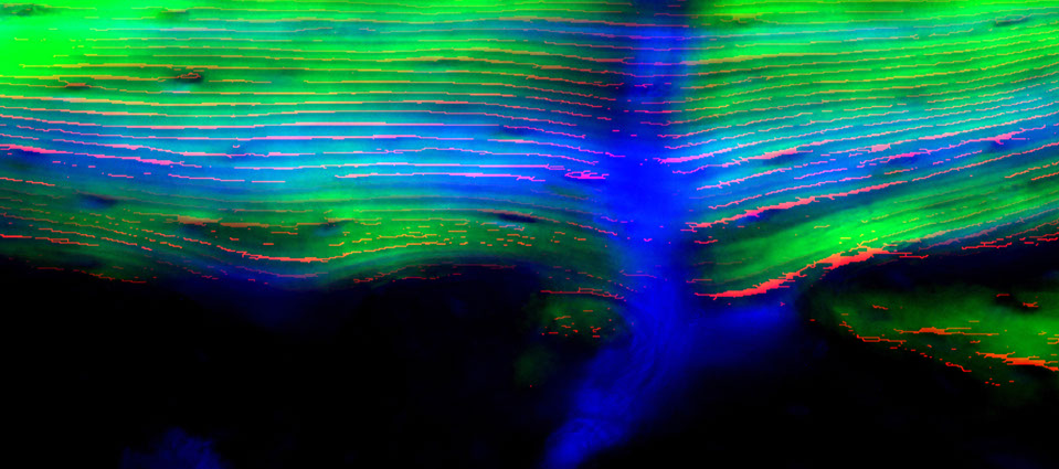


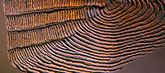


Rats in Space
How do changes in gravity from earth to outer space affect bone growth? This image shows changes to the bone of a growing rat that spent time on the space shuttle. Each horizontal line represents small changes to the bone over a 24-hour period.
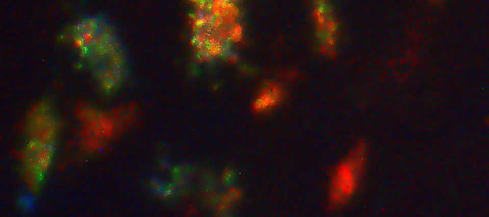
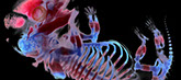
Mouse Embryo
A 16-day-old mouse embryo. The embryo
has been colored to reveal a flexible connective material known as cartilage (blue) and bone (red). Observations made from this image help scientists who are researching the evolution of the skeleton as well as treatments for skeletal disorders, cancer, and other diseases.
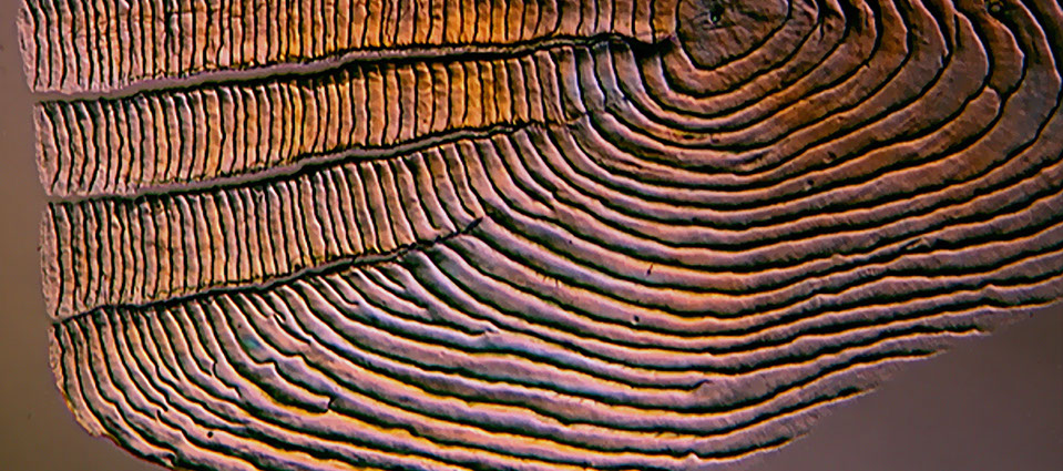
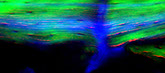
Fish in Space
This microscopic view shows the scale of a fish that spent time on the space shuttle. This image suggests that the fish adapted well to gravity changes in outer space: The growth of the scale, as illustrated by the lines radiating from its center, follows an orderly pattern, indicating that the scale continued to grow normally while in space.
Cave Bear
A close-up view of the bone of a cave bear who lived between 400,000 and 700,000 years ago in Spain. This microscopic image shows specific features of the skeletal remains of bears that were found with humans in the same caves. The red and blue colors highlight three-dimensional locations of features in the bone.
3 Million-Year-Old Bone
A microscopic view from a thigh bone of a three-million-year old early human. This image shows the bone cell spaces, corresponding to the the basic structural units of the bone. Comparing this image with images of bone from other early human species can help us to understand how humans have evolved over time.
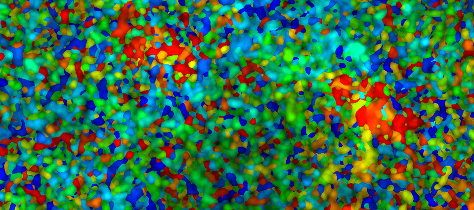
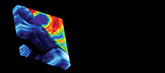
Thigh Bone
Researchers would like to better understand how a part of the bone that’s known as collagen grows. This view of a human thigh bone, which was re-created with three-dimensional computer imaging software, shows typical collagen growth patterns in 3D. The red, yellow, blue and green areas represent different growth patterns.
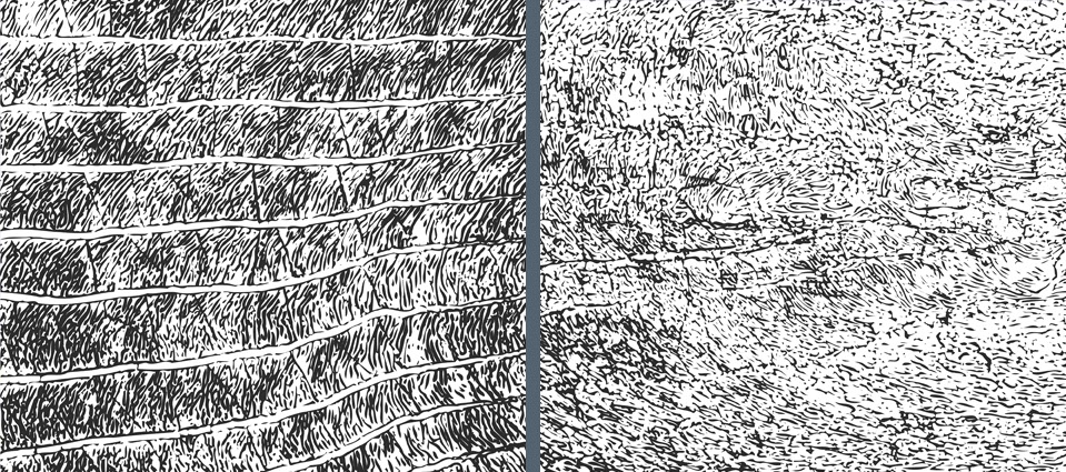
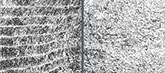
Fish scales
A microscopic view of two fish scales. The scales in one image have a predictable, rhythmic pattern; in the other, a chaotic and disturbed pattern. The two fish are the same species, except that the one with the chaotic pattern lived in a lake polluted by fallout from an accident at a nuclear power plant.
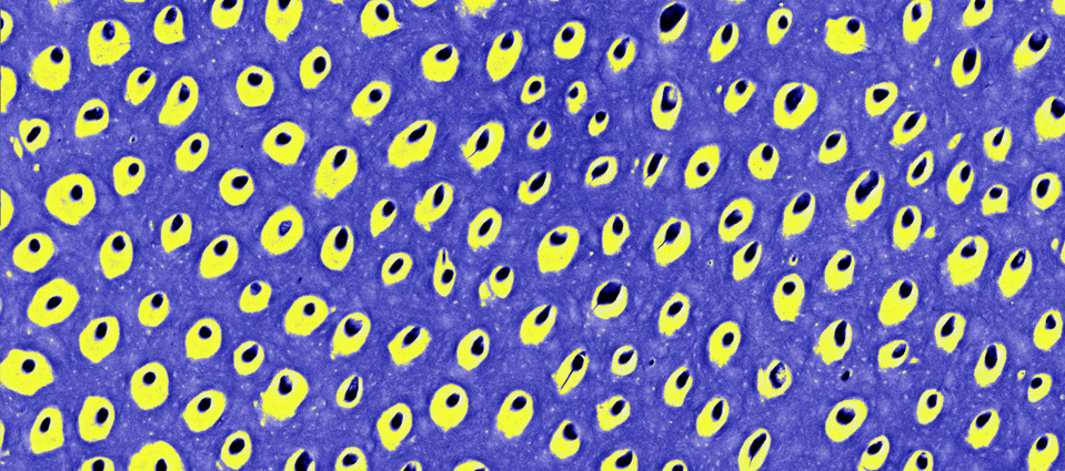
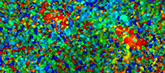
African Zebra
A microscopic view of the tooth surface of an African zebra. Black represents holes in the surface, yellow is the densest part of the tooth, and blue is the least dense section. Scientists study such images to better understand the effects of environmental change and disease on tooth and bone development.

Microscopes and Digital Imaging
The microscopes allow scientists to see tiny but important details in bones and teeth. And the digital imaging allows scientists to creatively alter colors to highlight these features.
Bone Growth
Did you know that your bones are growing and changing all the time?
In fact, your bones started growing even before you were born.


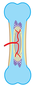
This is how your bones begin. First there is a cartilage model (blue) that’s slowly replaced by bone (yellow), starting in the shaft at the middle. Arteries (red) bring in blood to help things along.
This is how your bones first looked in the very earliest stage of life, before you were born, while you were first developing inside your mother’s body. During this period, tiny bones begin forming from a soft and flexible material called cartilage.
As time goes on, the bone grows bigger and bigger, and the cartilage begins to be replaced by the harder material we all recognize as bone. This is a gradual process that begins before you are born and continues after birth.
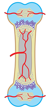
Slowly the shaft becomes all bone. Then cartilage is replaced by bone at the ends as well.
You’re still growing! Here’s how your bones look as they continue to get bigger and shed cartilage. This phase occurs at different times for different bones. It can begin as early as at birth, or as late as age 20.
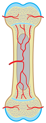
A bone that is almost done. Growth in the length of the bone continues at the thin layers of cartilage between the shaft and the ends, until you are fully grown.
At last, the growth process is coming to an end. This is how your bones look when they’ve grown to the point at which almost all of the cartilage has been replaced by bone. This stage begins as early as age 11, and in some cases, it can start as late as age 25.
When do you stop growing? It happens when you’re in your mid-twenties. Once all the cartilage has been replaced, you’ve grown to be as big as you’ll ever be.
+
Phase 1–3
+
Phase 4
+
Phase 5
Imprint/Legal Disclaimer
Publishers
Dr. Timothy G. Bromage
Hard Tissue Research Unit
Department of Biomaterials & Biomimetics
New York University College of Dentistry
345 East 24th Street
New York, NY 10010-4086
USA
Prof. Dr. Friedemann Schrenk
Senckenberg Gesellschaft für Naturforschung
Sektion Paläoanthropologie
Senckenberganlage 25
60325 Frankfurt
Deutschland
Disclaimer
Despite careful control of the contents, the paleobiomics team assumes no liability for the contents of external links. The operators are exclusively responsible for the contents of the linked sites.
Third party websites
Users may find advertising or other content on our site that links to the sites and services of our partners, suppliers, advertisers, sponsors, licensors and other third parties. We do not control the content or links that appear on these sites and are not responsible for the practices employed by websites linked to or from our site. In addition, these sites or services, including their content and links, may be constantly changing. These sites and services may have their own privacy policies and customer service policies. Browsing and interaction on any other website, including websites which have a link to our site, is subject to that website's own terms and policies.
Copyright
The contents of these Internet sites have been protected by copyright. Use, storage and/or reproduction of pictures and texts is not permitted as a matter of principle without express approval from the publishers. By using this site, you signify your acceptance of this policy and terms of service. If you do not agree to this policy, please do not use our site. Your continued use of the site following the posting of changes to this policy will be deemed as your acceptance of those changes.
Contact
If you have any questions, please contact us at mail(at)paleobiomics.org
x
Imprint/Legal Disclaimer
Publishers
Dr. Timothy G. Bromage
Hard Tissue Research Unit
Department of Biomaterials & Biomimetics
New York University College of Dentistry
345 East 24th Street
New York, NY 10010-4086
USA
Prof. Dr. Friedemann Schrenk
Senckenberg Gesellschaft für Naturforschung
Sektion Paläoanthropologie
Senckenberganlage 25
60325 Frankfurt
Deutschland
Disclaimer
Despite careful control of the contents, the paleobiomics team assumes no liability for the contents of external links. The operators are exclusively responsible for the contents of the linked sites.
Third party websites
Users may find advertising or other content on our site that links to the sites and services of our partners, suppliers, advertisers, sponsors, licensors and other third parties. We do not control the content or links that appear on these sites and are not responsible for the practices employed by websites linked to or from our site. In addition, these sites or services, including their content and links, may be constantly changing. These sites and services may have their own privacy policies and customer service policies. Browsing and interaction on any other website, including websites which have a link to our site, is subject to that website's own terms and policies.
Copyright
The contents of these Internet sites have been protected by copyright. Use, storage and/or reproduction of pictures and texts is not permitted as a matter of principle without express approval from the publishers. By using this site, you signify your acceptance of this policy and terms of service. If you do not agree to this policy, please do not use our site. Your continued use of the site following the posting of changes to this policy will be deemed as your acceptance of those changes.
Copyright
If you have any questions, please contact us at mail@paleobiomics.org
x
© 2014 Human Paleobiomics Hard Tissue Research Program by T.G. Bromage and F. Schrenk
© 2014 Human Paleobiomics Hard Tissue Research Program
Some related sites
Partners & Friends
+
+
+
+
+
+
+
+

